How Endodontists Use Advanced Microscopy to Save Teeth That General Dentists Cannot Treat
The Revolution in Root Canal Treatment
The landscape of dental care has undergone a remarkable transformation over the past few decades, with technological advancements enabling specialists to tackle cases that would have once resulted in inevitable tooth extraction. At the forefront of this evolution stands the dental operating microscope, a sophisticated piece of equipment that has fundamentally changed the capabilities of endodontic treatment. While general dentists provide excellent care for routine procedures, certain complex cases require the enhanced visualisation and precision that only an endodontist Buckinghamshire area or elsewhere can offer through advanced microscopy.
The difference between what can be achieved with the naked eye versus microscopic magnification is nothing short of extraordinary. General dental practitioners typically work with magnification levels of around 2.5x to 3.5x using loupes, whereas endodontic microscopes can provide magnification up to 25x or more. This dramatic increase in visual acuity allows specialists to identify and treat anatomical complexities that would otherwise remain hidden, ultimately saving teeth that might otherwise be deemed untreatable.
Understanding the Limitations of Conventional Dentistry
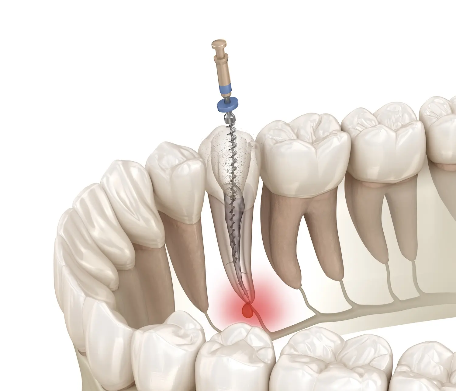
Root canal systems are remarkably intricate networks of canals, branches, and microscopic tributaries that weave through the tooth structure in patterns as unique as fingerprints. General dentists, despite their considerable skills and training, face inherent limitations when attempting to navigate these complex anatomical structures without specialist equipment. The human eye, even when aided by standard dental loupes, simply cannot detect the minute details that often determine treatment success or failure.
Research has demonstrated that microscope-enhanced endodontic treatment significantly improves success rates by enabling practitioners to locate additional canals, identify fractures, and remove separated instruments. These capabilities prove particularly valuable when treating teeth with calcified canals, unusual anatomy, or previous failed treatments. An endodontist with access to such technology can address cases that general practitioners must refer onwards, providing patients with genuine alternatives to extraction.
The Technical Advantages of Microscopic Endodontics
Illumination and Magnification Combined
Modern endodontic microscopes offer more than simple magnification; they incorporate coaxial illumination that projects light directly along the viewing axis. This configuration eliminates shadows within the canal system, revealing details that would remain obscured under conventional lighting conditions. The combination of magnification and superior illumination enables specialists to work with unprecedented precision, distinguishing between healthy tooth structure and diseased tissue with remarkable clarity.
Accessing Hidden Canal Systems
Teeth frequently contain more canals than standard anatomical textbooks suggest, with variations occurring in a significant percentage of cases. The ability to locate these additional canals proves crucial for treatment success, as any untreated canal can harbour bacteria and lead to persistent infection. Endodontists equipped with microscopes can identify these concealed pathways through careful examination of the pulp chamber floor, detecting subtle colour changes and anatomical landmarks that indicate canal orifices invisible to the unaided eye.
Complex Cases That Require Specialist Intervention
Certain clinical scenarios particularly benefit from microscopic treatment approaches. Retreatment cases, where previous root canal therapy has failed, present unique challenges that demand enhanced visualisation. The specialist must remove existing filling materials, locate missed canals, and address complications such as ledges or perforations. Similarly, teeth with calcified canals—where the natural canal space has mineralised over time—require microscopic guidance to safely navigate without damaging the tooth structure.
Separated instruments lodged within canal systems represent another category where microscopic intervention becomes invaluable. These fragments can often be retrieved or bypassed under magnification, whereas conventional approaches would necessitate extraction. Studies indicate that advanced microscopy enhances procedural outcomes across diverse endodontic scenarios, reinforcing the value of specialist referral for challenging cases.
The Distinction Between General Practice and Specialist Care
The decision to seek specialist endodontic care often depends on case complexity rather than any deficiency in general dental expertise. General practitioners rightfully recognise when a case exceeds the scope of conventional treatment and requires the advanced technology and focused training that specialists possess. This collaborative approach ensures patients receive appropriate care levels, with specialist endodontists offering distinct advantages for complicated scenarios whilst general dentists manage routine procedures effectively.
Preserving Natural Teeth Through Advanced Technology
The integration of microscopy into endodontic practice represents a significant advancement in conservative dentistry, enabling the preservation of natural teeth that previous generations would have lost. As technology continues to evolve, the gap between what can and cannot be treated widens in favour of tooth retention. For patients facing complex dental challenges, consulting an endodontist with access to advanced microscopic equipment may offer the difference between extraction and successful treatment, ultimately maintaining natural dentition and the quality of life that accompanies it.